

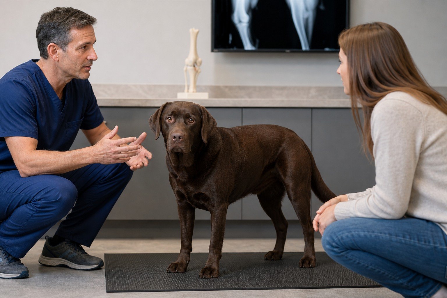
The cranial cruciate ligament helps stabilize the canine stifle, but canine cruciate disease is not always a healthy ligament suddenly torn by one misstep. Current evidence supports a multifactorial degenerative model for many spontaneous failures. Lameness, stiffness, difficulty rising, an altered sit, or reduced activity can suggest pain without identifying its source. Veterinary history, hands-on assessment, imaging when indicated, and patient-specific discussion are needed before medical, surgical, rehabilitation, or monitoring decisions.
Short answer
A sudden limp does not tell the whole ligament story
Cruciate disease may appear suddenly, gradually, or after an ordinary activity that makes an existing problem visible. ACVS describes a spectrum of pain and hindlimb dysfunction, while a scoping review supports a multifactorial degenerative process for many spontaneous failures. Neither onset pattern proves the diagnosis, mechanism, or best treatment. [1] [2]
- Record the exact pattern, timing, exposures, function, and recent changes.
- Separate population evidence from what can be concluded about one dog.
- Use the veterinary team to define evaluation, monitoring, and urgent-response steps.
Safety first
Acute non-weight-bearing lameness or severe pain needs prompt assessment
Contact a veterinarian promptly for a new or worsening hindlimb limp, swelling, pain, altered sitting, difficulty rising, or reduced activity. Seek urgent or emergency care after major trauma, for severe distress, inability to stand, dragging or knuckling paws, collapse, breathing difficulty, pale gums, or rapid deterioration.
- Do not use general information to delay care for severe, persistent, or worsening signs.
- Do not start, stop, combine, or calculate treatment from this article.
- Keep labels, dates, videos, and a concise observation timeline available for the veterinary team.
Veterinary note
This article is educational and does not diagnose, treat, prescribe, or replace care from a licensed veterinarian. Your dog’s history, examination, diet, medications, and current signs determine what is appropriate.
Understand the stifle and disease model
The cranial cruciate ligament limits abnormal forward movement and rotation within the stifle. Disease or rupture can produce instability, pain, altered loading, inflammation, and progressive joint change. The meniscus and other structures may also matter, but this article does not provide a home stability or meniscal test. [1]
Many canine cases differ from an acute traumatic ligament injury in a human athlete. Current literature describes complex contributions from ligament biology, inflammation, conformation, mechanics, genetics, body condition, and other factors. An ordinary turn or jump may coincide with onset without proving it was the sole cause. [2]
A gradual limp does not prove cruciate disease either. Hip, patella, spine, nerve, muscle, tendon, foot, fracture, infection, immune disease, or tumor can produce overlapping signs. The veterinary examination must keep alternatives visible.
Record what the dog is doing differently
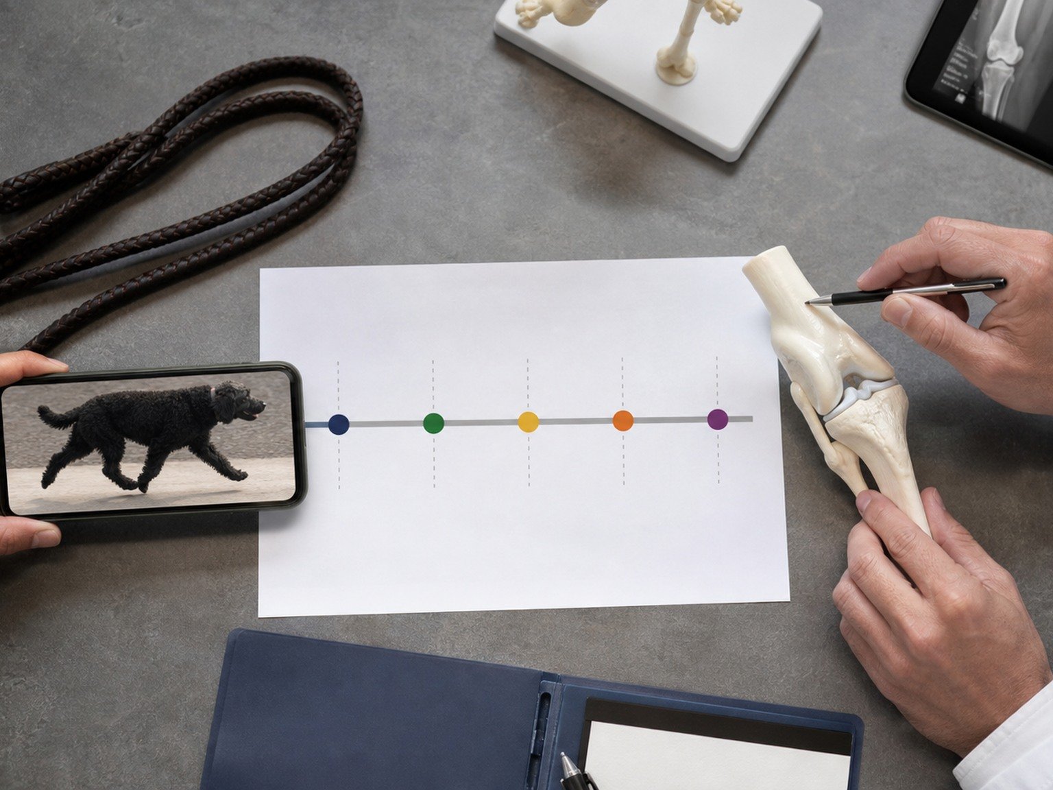
Owners may notice variable weight bearing, stiffness after rest, difficulty rising, reluctance to jump, reduced play, an unusual sit, or changes on stairs. Record the affected side only as an observation; compensatory loading can make the opposite limb look different too.
Capture onset, progression, surface, activity, rest response, medications, previous lameness, and a few safe walking videos from consistent angles. Do not repeatedly provoke running, turning, stairs, or jumping to demonstrate the problem. Preserve comfort while collecting only naturally occurring evidence.
AAHA pain guidance emphasizes owner input, reproducible assessment, and reassessment. A clinic visit is a snapshot, so daily-function records can show what is missed, but they do not replace palpation, gait assessment, neurologic screening, and imaging decisions. [4]
Follow the veterinary assessment pathway
The clinician considers onset, trauma, prior episodes, activity, age, body condition, medications, concurrent disease, and caregiver goals. Orthopedic and neurologic assessment helps localize the problem. Sedation, radiographs, or referral may be used when needed for a reliable, safe evaluation. [1]
Radiographs do not show the ligament like a simple photograph, but they can show joint changes and help evaluate alternatives. Findings are interpreted with the examination. Do not use an image report, drawer demonstration online, or the sound of a click to diagnose the dog at home.
Risk-factor reviews identify associations involving joint disease, body weight, breed, age, and conformation, but evidence strength varies and interactions are incomplete. These factors cannot predict the diagnosis, second-limb course, or outcome for an individual. [3]
Prepare for a patient-specific decision
Management discussions may include pain control, activity restriction, surgery, medical management, weight and nutrition, rehabilitation, environmental support, and follow-up. The appropriate combination depends on instability, size, activity, age, conformation, meniscal concern, other disease, caregiver capacity, and clinical findings.
This guide does not compare procedures, publish success percentages, or prescribe rehabilitation. Ask what each option is intended to change, what uncertainty remains, what restrictions apply, how complications are monitored, and whether a board-certified veterinary surgeon or rehabilitation professional should be involved.
Clarify written instructions for medication, movement, incision care if applicable, emergency signs, recheck, and return to activity. Do not give human pain medicine, use leftovers, or improvise exercises. A good plan changes when function, pain, healing, or circumstances change.
Prepare for a focused veterinary conversation
Bring a concise timeline, short natural-movement or symptom videos when safe, the exact names and photographs of every food, treat, medication, and supplement label, and notes about appetite, water intake, stool, sleep, activity, comfort, and behavior. Include recent injuries, travel, boarding, diet changes, missed medication, and previous test results. A complete record helps the veterinary team separate a repeatable pattern from a single impression.
Decide in advance what you need from the visit: an urgency decision, a diagnosis plan, a nutrition review, a pain or mobility assessment, or a monitored trial. Ask what result would change the plan and what finding would rule an option out. This keeps research and product information in the right role. Evidence can shape questions and expectations, but it cannot determine what is safe for an individual dog without the history and examination.
Owner tool
Prepare for a cruciate consultation
On a phone, swipe across the table to see every column.
| Checkpoint | Record | Why it helps |
|---|---|---|
| Onset | Sudden or gradual, activity, side | Frames mechanism |
| Function | Rise, walk, sit, stairs, play | Shows impact |
| History | Prior limp, drugs, disease, weight | Adds context |
| Plan | Options, restrictions, recheck | Supports decisions |
Better questions, calmer next steps
Questions to ask your veterinarian
- Which findings localize the problem to the stifle?
- What alternatives are still being considered?
- Would imaging, sedation, or referral change the plan?
- What outcome and complication signals should I track?
- Who coordinates pain care and rehabilitation?
FAQ
Did one jump tear a healthy ligament?
Not necessarily. Many spontaneous failures have a multifactorial degenerative background.
Can I test the knee at home?
No. Stability assessment belongs to the veterinary examination.
Do radiographs show the ligament?
They mainly support joint assessment and alternatives when interpreted with the exam.
Is surgery always the same?
No. Options and suitability are patient-specific.
Should I start exercises now?
Only follow written instructions from the treating veterinary team.
Sources
- American College of Veterinary Surgeons: Cranial Cruciate Ligament Disease. Anatomy, signs, diagnosis, and individualized decisions.
- Animals: Etiopathogenesis of Canine Cruciate Ligament Disease. Multifactorial degenerative disease model.
- Frontiers in Veterinary Science: Risk Factors for Canine Osteoarthritis. Risk evidence and uncertainty.
- American Animal Hospital Association: 2022 Pain Management Guidelines. Owner input and patient-specific reassessment.
